Wednesday, 12 June 2013

Parasites and Networks - Food Webs, Epidemiology

When we think about how parasites fit into nature - their close interaction with their hosts, their reliance on specific hosts in a specific sequence, their ubiquitous presence in the environment - a network (or web) context makes sense. Recently, two articles  have come to my attention; they represent parasites in webs in similar ways but for different reasons.

Parasites Affect Food Webs

This food web for a mosquito would be much
more complex - and of much more relevance
to humans - if it included parasites.
Illustration by Tyler Rubley. CC BY-SA 3.0


The first is "Parasites Affect Food Web Structure Primarily Through Increased Diversity and Complexity." by J. Dunne and co-authors (PLoS Biol 11(6): e1001579), published on June 11, 2013. These researchers added parasites to aquatic food webs and asked whether the changes that resulted (a much more complex food web) could have been caused by the addition of any large group of organisms, or whether food web structure was altered by parasites in unique ways.

Beyond the central findings of that study, the article raised (or re-raised) a question for me: if we know that parasites are an important component of an ecosystem, if we know that they affect food webs in both generic and unique ways, why do they never appear on endangered species lists? For every endangered species, there should be a list of parasites that depend on that host species for survival. And why do conservationists routinely rid endangered animals in captivity of their parasites, knowing that if that species ever returns to the wild, it will do so without its specialist parasites, potentially to its detriment?

Parasites Predict Disease


The other paper is "Centrality in Primate–parasite Networks Reveals the Potential for the Transmission of Emerging Infectious Diseases to Humans" by José María Gómez and co-authors , published in PNAS (110:19 2013).  Like the paper by Dunne et al, this research builds a food web, but this web has parasites as the main consumers - a network of nonhuman primates that share parasites (or that are consumed by the same parasites, to look at it from the parasites' viewpoint).

The point is to examine whether this web can predict where diseases of primates are likely to jump to humans. The authors "found that primate species having higher values of centrality in the primate–parasite network harbored more parasites identified as EIDs [emerging infectious diseases] in humans and had parasite communities more similar to those found in humans." These species, then, are more likely to be the source of emerging infectious diseases in humans. Another good reason to include parasites in food webs.

Food webs tell us where parasites fit, and I think that is something we really need to understand, for our own sake and for theirs.

Friday, 22 March 2013

Disseminated Strongyloidiasis - Interesting Things to Note

A recent case of disseminated strongyloidiasis, reported by the New England Journal of Medicine highlighted a couple of intriguing features of this catastrophic disease: in the absence of an effective immune response, worms can mature in the lungs as well as the intestine, and, there's something special about corticosteroid drugs that makes an unfortunate success of the worm.

Adult Strongyloides stercoralis in the Lungs


The case report of disseminated strongyloidiasis by Schroeder and Banaei describes adult worms, both rhabditiform and filarifom larvae, and ova containing active larvae in a tracheal aspirate. A similar case is reported by Bava et al. Typically, you'd find only filariform larvae in the lungs, and adults are almost never seen, even in the stool, because the adult females spend their time migrating through the tissues of the intestinal lining (and there are no parasitic males).


A larva of Strongyloides stercoralis. At a later stage, this larva would
be capable of penetrating and migrating through tissues like skin, or the
lining of the intestine. Image: CDC


I assumed that the adults found in this case had actually matured in the lungs rather than migrating there from the intestine. A 2004 paper in Clinical Microbiology Reviews agrees. Keiser and Nutman write “...findings suggest that filariform larvae develop into adults in the lungs... This hypothesis is supported by... autopsy studies showing adult worms in lung tissue.” In this scenario, the parasite could be multiplying very rapidly with new worms originating not only in the intestine, but in the lungs as well. One can only imagine the numbers of parasites that could be present within a short period of time.

Corticosteroids and Strongyloides stercoralis


We know that corticosteroids can initiate disseminated strongyloidiasis. But do they just give the worms a green light by suppressing immune response, or do they actually favor the parasite? Corticosteroids prevent production of eosinophils and cause the rapid destruction of eosinophils that already exist; these cells are part of the body's immune response to parasites. But it's thought that corticosteroids actually contribute to the success of S. stercoralis in another way. Gary Simon writes in Medical Parasitology that “they may stimulate female worms to increase larval output and promote molting of rhabditiform larvae into the invasive filariform larvae.”

Eosinophils in the blood are part of the
immune response to parasites. Image by
Iceclanl. (cropped)  CC BY-SA 3.0

So it looks like giving corticosteroids to a patient with S. stercoralis expands the “territory” in which the parasite can reproduce, hobbles the immune system's attempts to control it, and boosts the worms' fecundity and maturation. Given all of this, it's easy to see why it might be difficult to save a patient suffering from disseminated strongyloidiasis, unless the problem is discovered quickly. It also sheds some light on why other types of immunosuppression are relatively less catastrophic.


Sources


Bava  BAJ, Cecilia D et al. “Adult Female of Strongyloides stercoralis in Respiratory Secretions.”, Asian Pacific Journal of Tropical Biomedicine 3:4, April 2013, Pages 311–313

Keiser PB, and Nutman TB. “Strongyloides stercoralis in the Immunocompromised Population.” Clinical Microbiology Reviews. 17:1, January 2004, 208–217.doi:10.1128/CMR.17.1.208-217.2004

Castelletto ML, Massey HC Jr et al. "Morphogenesis of Strongyloides stercoralis Infective Larvae Requires the DAF-16 Ortholog FKTF-1." , PLoS Pathogens 5(4): e1000370. doi:10.1371/journal.ppat.1000370

Schroeder L, and Banaei N. “Strongyloides stercoralis Embryonated Ova in the Lung.” New England Journal of Medicine: March 21, 2013; 368:e15 http://www.nejm.org/doi/full/10.1056/NEJMicm1204579

Simon, G. “Strongyloidiasis.” In: Medical Parasitology. Satoskar AR et al eds. Austin: Landes Bioscience; 2009, pg 31

Tuesday, 19 March 2013

Beauveria bassiana - A Fungus That Kills Bed Bugs

Beauveria bassiana is a fungus that is well known for killing insects. Spores of B. bassiana adhere to the cuticle (the outer protective covering) of the insect, begin to grow, and work their way through to the inner tissues. There the fungal growth continues, taking nutrients from the host's body until the insect dies. Because B. bassiana is lethal to many insects, including their larvae, it has been grown and distributed commercially for use in agricultural control of insect pests. It makes sense to wonder whether it could be used to control bed bugs as well.
Beauveria bassiana is grown for control of agricultural
pest insects. Image courtesy of Keith Weller.

Beauveria bassiana Kills Bed Bugs


A study by Alexis Barbarin et al tested B. bassiana against bed bugs, and the results indicate that the fungus is lethal to the pests: not only does it kill virtually all bugs that come in contact with it, infected bugs can carry it back to daytime hiding places and pass it on to other bugs that have not been otherwise exposed. Barbarin el al propose that B. bassiana might rid a bed bug infested building of its bugs.

Bed Bug Traps Using Beauveria bassiana


In their study, Barbarin et al exposed bed bugs to a mixture of oil and fungal spores on various surfaces, and found that jersey knit cotton transmitted the infection most effectively. Though further research is required, they propose that a fabric bed skirt impregnated with B. bassiana spores might be an efficient means of infecting a resident bedbug population. Presumably any trap designed so that all bedbugs climbing onto or leaving the bed would have to pass through it could be used to infect them with the fungus.

Is Beauveria bassiana Safe for Humans?


Beauveria bassiana is generally regarded as safe for humans and it's already being used for insect control applications all over the world without dire consequences for human health. This fungus is already naturally occurring in the environment. However, there's reason to be cautious with this approach. A study that tested fungi isolated from poultry barns found that B. bassiana has several virulence factors that potentially “increase [its] survival, growth, and propagation... in animal tissue.” Authors Taira el al comment, quite correctly, that otherwise harmless fungi can cause serious infections in people whose immune systems are already compromised. Such people include AIDS patients and organ donor recipients among others.

These grasshoppers were killed by Beauveria bassiana.
Fungal growth is visible on the insects' remains.
Image courtesy of Stefan Jaronski.

Cases of both deep tissue infection and skin infection caused by B. bassiana have been reported in the medical literature (Figueira et al). The possibility of skin infection, in particular, prompts second thoughts. Beauvaria bassiana does not wipe out bed bugs on contact: it takes time for the infection to kill. Therefore, bugs that have contacted the fungus will still visit the sleeping host to feed. And while feeding, they will create a break in the skin, often with severe irritation resulting, and possibly introduce fungal spores. This does not seem like a good plan.

Beyond the possibility of skin infection arising from contact with the bugs, a spore impregnated bed skirt would presumably contaminate a living space with fungal spores pretty thoroughly, and fungal spores are as hardy and as hard to eliminate as bed bugs are. Under the right conditions, they could remain viable for a very long time, possibly protecting against reinfestation by bed bugs, but also a potential hazard for the immunocompromised occupant of, or visitor to, the space.

Sources


Barbarin AM, Jenkins NE et al. “A Preliminary Evaluation of the Potential of Beauveria bassiana for Bed Bug Control.” Journal of Invertebrate Pathology 111 (2012) 82–85

Figueira L, Pinheiro D et al. “Beauveria bassiana Keratitis in Bullous Keratopathy: Antifungal Sensitivity Testing and Management.” European Journal of Ophthalmology 22:5 (2012) 814-818

Taira CL, Marcondes NR et al. “Virulence Potential of Filamentous Fungi Isolated From Poultry Barns in Cascavel, Paraná, Brazil.” Brazilian Journal of Pharmaceutical Sciences 47:1 Jan./Mar. 2011


Tuesday, 12 February 2013

Every Living Thing: A Book Review

Every Living Thing: Man's Obsessive Quest to Catalog Life, from Nanobacteria to New Monkeys
by Rob Dunn
Harper 2010 ISBN 978-o-06-143031-2

“The biological world does not revolve around us... life is smaller than we imagined, ...we are a smaller part of life than we imagined,” and we occupy a “marginal position in the biological universe” (Every Living Thing p. 59 - 60). With Rob Dunn, you know where we stand. But from that marginal position, he's written a fascinating book about the life we share our planet with, and the key people who've helped us learn what we know about that life.

I found Every Living Thing to be a eye-opener in two ways. First, it reminded me forcefully that in spite of all that we know, we still don't know much. For someone who once believed (albeit a very long time ago) that all the answers were out there – you just had to go to the right library and ask the right question – it was both bewildering and liberating to realize that what we don't know is actually in the majority. To contemplate the vast quantity of scientific knowledge stored in academic libraries and realize that it is only the beginning is truly shocking.

'Animalcules' illustrated by
Anton van Leeuwenhoek; c1795.
Second, Dunn has illuminated the lives and accomplishments of some people who are frequently mentioned only briefly, even in the most interesting science books. I knew that Antonie van Leeuwenhoek discovered Giardia lamblia when he observed it in his own stool by looking through his own glass microscope lenses. I did not know that he was the first person known to observe microscopic life of any kind; that he discovered two entire kingdoms of life; that he was a family man with five children. I didn't know that two scientists who changed the world in very different ways – Lynn Margulis and Carl Sagan – were actually married to each other at one time. The book is full of surprises like that.

If there is anything that makes Every Living Thing a challenge, it's frequent jumps through time and space, and from third to first person narrative. Far from being chronological, the narrative draws unexpected connections throughout. The reader who is fully engaged and paying close attention will be fine. Others may feel suddenly disoriented and have to retrace their steps to find out how they got from 1960s California to 1848 Brazil; from first person contemplation of becoming a father, to endosymbiogenesis. The connections are relevant; they clarify and enlarge the picture, but it is, at times, quite a ride.

Dunn has a talent for explaining complex science in a way that the general reader can understand and he combines this talent with great storytelling. This grounds the people he writes about in a context of everyday life we can all relate to, and results in a very interesting and relevant science book. It's easy to imagine that Every Living Thing will end up in the reference section of a lot of personal libraries, including mine.


At one time, I was a great fan of Stephen Jay Gould, but today I'd have to say that both in print and online, Rob Dunn is my favourite science writer. I hope this strong beginning is just the first of many fascinating science books.

Tuesday, 5 February 2013

Was Joshua the Hero Of Jericho? (Schistosoma and Jericho)

The Jordan Valley near Jericho.
From the OSU Special Collections
and Archives.
Reform Judaism Magazine has published an adapted excerpt from my book Parasites: Tales of Humanity's Most Unwelcome Guests. The excerpt is a shortened version of my retelling of the story of Rahab, Joshua, and the Battle of Jericho, with the blood fluke Schistosoma hematobium as a central character.
 
Reform Judaism Magazine is an interesting publication. The online version features a number of articles that would appeal to any reader: titles that caught my eye include "Were the Jews Slaves in Egypt?," "The Divinity of Dementia," and "A Paragraph that Changed History" ("Why Jesus' last supper could not have been a Passover meal..."). Fascinating.

It's my pleasure to be published in this magazine. You can read Was Joshua the Hero of Jericho? on the internet.


Thursday, 31 January 2013

Echinococcus multilocularis Liver Cysts in Dogs

Infected foxes spread Echinococcus
multilocularis to dogs.
Image by Andy Potter; CC BY-SA 2.0.
As any good parasitology text will tell you, liver cysts caused by Echinococcus multilocularis typically occur in rodents: animals like voles, lemmings, and mice. The disease is called alveolar echinococcosis, or alveolar hydatid disease, and it occasionally occurs in people too, if eggs of the tapeworm are accidentally swallowed.

Echinococcus multilocularis liver cysts in dogs


The liver cyst caused by E. multilocularis is a larval stage - a stage that multiplies asexually in the cyst. The adult stage of the parasite is found in canids, members of the dog family: arctic foxes, red foxes, jackals, coyotes, domestic dogs. Thus, the 2009 discovery of a liver cyst in a domestic dog in British Columbia, Canada (Jenkins et al.), is puzzling and alarming.

This scenario is not actually so bizarre. Taenia solium, or pork tapeworm, has a similar story: humans normally host the adult tapeworm in the intestine after consuming the larval cysticercus in undercooked pork. But if a human swallows the tapeworm egg instead, the eggs hatches and the larva moves into the tissues – sometimes the brain – and forms a cysticercus.

When humans have T. solium cysticerci in their tissues, the disease is called cysticercosis – or, in the brain, neurocysticercosis – and this can be much worse than having the tapeworm in the intestine. Clearly, in a dog, a liver cyst that can grow large enough to compromise liver function, and spread to other parts of the body, is worse than hosting a tapeworm in the intestine as well.

In cysticercosis in humans, and presumably alveolar echinococcosis in dogs, the tissue cysts often originate from the hosts own intestinal tapeworms. In humans, it’s poor hygiene and hand to mouth transmission. In dogs, it’s grooming – licking eggs off soiled fur. This raises the question: have dogs always frequently had the liver cysts when they had the worms or has something changed?

Echinococcus multilocularis spreading to new places


Jenkins et al. remark that “compared with native North American strains, European strains of Echinococcus multilocularis appear to have greater potential to cause alveolar hydatid disease (AHD) in humans.” The strain found in British Columbia was a European strain; perhaps they have greater potential to cause AHD in dogs as well. Do we know?

The British Columbia dog apparently did not have the adult tapeworm in its intestine and the authors speculate that the parasite may have been introduced by an imported infected dog. This, too, is alarming. It brings to mind my discussion of E. multilocularis in Parasites: Tales of Humanity’s Most Unwelcome Guests, in which I relate the identification of Echinococcus multilocularis in wild canids smuggled into the Eastern US for fox hunting.

If one imported dog can introduce the worm to British Columbia, what is the likelihood that many illegally translocated infected canids have not spread the worm as well? Is alveolar echinococcosis simmering in rodents, dogs, and people in the eastern United States?

Sources


Jenkins EJ, Peregrine AS, Hill JE, Somers C, Gesy K, Barnes B, et al. Detection of European strain of Echinococcus multilocularis in North America [letter]. Emerg Infect Dis [serial on the internet]. 2012 June.

Roberts, Larry S., and John Janovy Jr. Gerald D. Schmidt & Larry S. Roberts’ Foundations of Parasitology 8th ed. Boston: McGraw Hill, 2009. Pg 354-5.

Thursday, 24 January 2013

Echinococcus multilocularis and Alveolar Echinococcosis

The tapeworm Echinococcus multilocularis normally spends its adult life in the intestine of a fox, usually an arctic or red fox. The fox acquires the worm by eating an infected rodent. In turn, the fox passes eggs in its feces, which rodents accidentally eat. In the intermediate host (the rodent or, sometimes, a human) the parasite occupies the liver rather than the intestine.

Alveolar echinococcosis


The adult Echinococcus multilocularis tapeworm is very
small. The head (or scolex) of this one is to the right.
Image: CDC
In the liver, the larva forms an alveolar cyst, a cyst composed of thin-walled chambers that multiply until the parasitic growth looks a bit like a mass of bubbles. These bubbles may break away and be carried to other parts of the body where they continue to grow. This is one reason why the alveolar cyst of E. multilocularis is often likened to a malignant tumor.

This is a nasty parasite and published accounts of the course of the disease in humans are rather horrifying. It spreads and is difficult to treat. It’s often fatal. This one, you never want to get. The literature and the media, meanwhile, lend the distinct impression that E. multilocualris is spreading and cases of alveolar echinococcosis in humans are becoming more common.

Distribution of Echinococcus multilocularis


A map in a 1984 parasitology text showing the global geographic distribution of E. multilocularis suggests that the worm has not broadened its horizons much in the last thirty years. In 1984, its range included most of Asia north of 40º latitude, Central and Eastern Europe, northern Canada, coastal Alaska, and a patch right in the middle of North America bisected by the Canada – US border and by 100º longitude. Today the distribution is marginally wider: more of Central Europe, parts of Western Europe, and the patch in the middle of North America has grown as well. Sporadic cases appear in other far flung regions: northern Africa, British Columbia in Canada.

So E. multilocularis is perhaps creeping rather than sweeping around the northern hemisphere. In truth, human infections are still extremely rare. Alveolar echinococcosisis, however, is “emerging” in humans in European locations, and there’s concern that it might do the same in parts of North America (most North American cases in the past have been in Alaska). In a recent paper in PLOS Neglected Tropical Diseases, Nahorski  and others report that, in Poland, only two cases were known prior to 1980, compared with 121 cases diagnosed between 1990 and 2011.

Why is alveolar echinococcosis increasing?


Better diagnosis is certainly one reason for the increase, and Nahorski et al feel that many undiagnosed cases remain. They point to another possible cause however - a boom in the urban fox population. “In Poland,” they write, “the fox population increased from 67 000 in 1995 to 220 000 in 2006.” That’s a very significant increase, and many of the human cases came from provinces where the worm is especially common in foxes. The data led the authors to conclude that infected domestic dogs and cats are also important sources of the disease.

In order for a domestic animal to acquire the intestinal worm, the animal would have to eat an infected rodent. Cats, of course, are hunters, but according to the European Scientific Council Companion Animal Parasites (ESCCAP): “Cats, in contrast to dogs, are epidemiologically insignificant as sources of egg output as they are poor hosts for this worm.” Dogs are a different matter: they are good E. multilocularis hosts, and many domestic dogs do hunt. Many do not of course, especially urban dogs. One would have to know one’s dog to judge the relative likelihood that it would ever have E. multilocularis eggs in its feces, or on its fur.
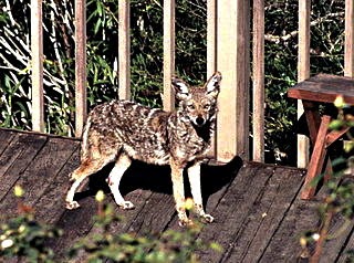
Urban coyotes have little fear of people.
Image by Steve Jurvetson, Menlo Pk, USA;
 CC by 2.0
Do foxes live in North American cities like they do in Europe? It’s true we don’t often read or hear about this, but they do: the city of Mississauga is one municipality that has addressed the issue of foxes in the city. And if those urban foxes have E. multilocularis, that could contribute to the emergence of alveolar echinococcosis in humans. But in North America, we have another canid that likes to live in cities, and it, too, can harbour E. multilocularis: the coyote. A study of urban coyotes in Chicago concluded that there are hundreds, perhaps thousands of coyotes living in that city, and those researchers believe that “the results likely apply to most major metropolitan areas in North America.”

So, while there's no reason to be paranoid about this rare disease, awareness of it isn’t a bad thing. All the usual advice still applies: wash your hands often, keep your dog close, and provide your dog with good veterinary care including screening for intestinal parasites.

Resources


Beaver, Paul C., Jung, Rodney C., and Eddie W. Cupp. Clinical Parasitology 9th ed. Philadelphia: Lea & Febiger, 1984. Pg 534.

City of Mississauga. Animal Services: "Foxes." 1995-2013

ESCCAP. “Worm Control in Dogs and Cats: ESCCAP Guideline 01 Second Edition.” September 2010

Nahorski WL, Knap JP, Pawłowski ZS, Krawczyk M, Polański J, et al. "Human Alveolar Echinococcosis in Poland: 1990–2011." PLoS Negl Trop Dis 2013; 7(1): e1986. doi:10.1371/journal.pntd.0001986

Wagner, Holly. “On the Loose: Urban Coyotes Thrive in North American Cities.” Ohio State Research News. Last Updated 2005.