Showing posts with label Parasites. Show all posts

Monday, 31 March 2014

Neospora caninum and Neosporosis

The sign on this public footpath in the UK reminds dog
owners to pick up after their pets. Cattle in the area have
aborted due to Neospora caninum.  Image by Peter Barr
 CC BY-SA 2.0
Neospora caninum, a parasite of dogs, causes abortion in cattle

Neospora caninum is a coccidian parasite of domestic dogs but it causes big problems for the cattle industry. We’re still learning about N. caninum and neosporosis.

In August 2007, dog owners in Somerset, UK were asked to be especially vigilant about picking up after their dogs. The reason was a spike in the number of cattle aborting fetuses in the area—some of the cattle tested positive for a parasite, Neosporum caninum, carried by domestic dogs.

The parasitic disease called neosporosis was first recognized in domestic dogs in Norway in the 1980s. It’s now known that the parasite is present in dogs, cattle, and other animals worldwide. A coccidian, it’s related to Toxoplasma gondii and Cryptosporidium, well known causes of parasitic disease in humans.

Life Cycle of Neospora caninum


The life cycle of N. caninum in dogs is very similar to that of T. gondii in cats. An infected dog has parasites multiplying sexually in the intestine and the infective oocyst is passed in the dog’s feces. Meanwhile, parasites are also multiplying asexually in other tissues.

Harbouring the sexual form of N. caninum in the intestine makes domestic dogs the definitive host of the parasite. It’s not known whether other animals are capable of serving as a definitive host, but wild dogs, such as foxes, wolves, coyotes etc. may do so.

Animals other than dogs that ingest either oocysts in dog feces or animal tissue in which the parasite is present become intermediate hosts – they have only the asexual stage, multiplying in the tissue. Thus, dogs pass on the parasite in their feces, in their tissue if they are eaten by other animals, or to an unborn fetus. No animals other than dogs have been known to spread the parasite in feces.

Neosporosis in Dogs


Neospora caninum infects domestic dogs worldwide with varying prevalence. Studies testing dogs for antibodies to the parasite suggest that more than 30% of dogs are infected in some areas, with the highest numbers in South American countries and in rural dogs, especially those living on cattle farms.

Most infected dogs have no symptoms. When symptoms occur, neosporosis is most severe in newborn puppies, infected during gestation when the parasites move from the bitch’s tissues to the fetus. Puppies suffer paralysis, particularly of the hind legs, and often do not survive. Adult dogs may suffer from an illness similar to toxoplasmosis in cats, or they may develop dermatitis.

Neosporisis in Cattle


Like dogs, cattle everywhere harbour N. caninum, and most show no signs of it. In some herds, close to 90% of cattle are infected and the parasite is thought to account for more than 40% of abortions – a significant cause of economic loss for cattle farmers. Many infected fetuses and calves appear normal, however, and it is still unclear what factors cause or prevent disease symptoms.

In cattle, N. caninum is transmitted only from a pregnant cow to her fetus—the parasite does not pass between cows in a herd. Some cows, then, must acquire the parasite from dogs, consuming oocysts while grazing where dogs have defecated. It’s easy to imagine how farm dogs and livestock (sheep, goats, and horses can also be infected) may have increased the prevalence of the parasite, with dogs eating the remains of aborted young, becoming infected, and then passing infective oocysts in feces deposited where livestock graze. Calves born without symptoms, meanwhile, pass the parasite on to their own young.

Whether the abortions occurring in Somerset in England resulted from infected dogs defecating on cattle farms, or whether they came from silent infections already present in the cattle remains unknown; however, picking up after your dog is always good practice, and will lessen the risk of spreading not only neosporosis, but other diseases as well.

Sources:


Foundations of Parasitology 8th Ed. Roberts, Larry S. and John Janovy Jr. Boston: McGraw Hill, 2009.

Review of Neospora caninum and Neosporosis in Animals. Dubey, J. P. The Korean Journal of Parasitology 41:1 Mar 2003, 1-16.

Wednesday, 22 January 2014

Elephantiasis and Wolbachia: The Bacteria Behind the Worms

Horrific parasites sometimes turn out to be mere vessels.

Filarial worms, nematodes that are transmitted by insects and live in the tissues, cause horrible disfigurement, disability, blindness. The common names for the diseases they cause are descriptive and apt: elephantiasis, and river blindness.

Wucheria bancrofti, and Brugia malayi nematode worms live in human lymph vessels. Prolonged presence of the worms there often results in the affected part – often a limb or the scrotum - becoming grossly enlarged and deformed. Microfilaria, the worm’s young, circulate in the blood.

Elephantiasis results in almost
unbelievable disfigurement of
various parts of the body.
Imagine living with this.
Tropenmuseum of the Royal
Tropical Institute (KIT)
CC BY-SA 3.0

Onchocerca volvulus adults live together in nodules in the skin. Their young migrate through the skin and, over the long term, can cause discoloration, aging, and sagging of the skin. Worse, microfilariae in the eyes cause blindness.

For a long time, we thought that the worms themselves, and the microfilariae, caused these terrible responses to infection – that the battle between the human body and the worms caused blindness and disfigurement. But now it’s becoming clear that the real culprit is not the worms, but a type of bacteria living inside the worms. The problem is Wolbachia pipientis.

Wolbachia and Filarial Worms


Wolbachia is best known for living in insects, where it is typically parasitic and sometimes causes fatal disease. In nematodes, the relationship is different. In nematodes, Wolbachia is a symbiont: the bacterium actually provides nutrients to the parasitic worm that the worm can’t otherwise get, and without the Wolbachia, the worm can’t survive.

Researchers now have compelling evidence that what the human immune system responds to is actually the Wolbachia, not the filariae or the microfilariae. It’s the Wolbachia that causes elephantiasis and river blindness.

Wolbachia, Filarial Nematodes’ Achilles Heel


Treatment of the diseases caused by filarial nematodes has proven difficult for many reasons, not the least of which is that few drugs kill the adult worms. The discovery of their reliance on Wolbachia reveals a weakness that may be the worm’s undoing: instead of finding a drug to kill the worms, we can use a drug to kill the Wolbachia.

Ironically, if it’s the Wolbachia that’s actually the culprit, the worms end up as collateral damage.

Tuesday, 7 January 2014

Schistosomes: The Snail, the Prawn, and the Dam

It’s a story that has played out in Africa all too often. A river is dammed to control flooding, or for hydroelectric power, or to create a water reservoir in case of drought (or all of the above at once). The dam, of course, creates a large lake and lots of good habitat along the shore for aquatic plants, which in turn support a large snail population.

Schistosome Life Cycle


People come to live near the water’s edge, to use the water and to fish. Generally, human contact with water is high, particularly for children and fishers. If schistosomes are present, along with the right snail species, escalating levels of schistosomiasis soon follow. This is how it works:
  • People infected with schistosomes pass the eggs in urine or feces, and if those eggs find their way into fresh water, they hatch.
  • The emerging larva seeks out an aquatic snail – only the right snail will do – and penetrates the snail, after which it multiplies asexually in the snail’s tissues.
  • Larvae leave the snail host and, if a human enters the water, penetrate human skin, maturing to adult worms that reside in the tiny blood vessels around the bladder or intestine.
  • Adult worms produce eggs which are passed in the urine or feces, and so on.

It makes perfect sense that water resource projects can result in rapidly increasing incidence of schistosomiasis because they bring together the two hosts of the parasites – the human and the snail – in circumstances ideal for transmission. We hardly need a more complicated explanation. It seems, however, that there’s at least one other factor that’s been left out: the African river prawn.

River Prawns and Schistosomes

River prawns are not small!
This is Macrobrachium carcinus (molt).
Clinton & Charles Robertson. CC BY 2.0

Fascinatingly, the adult river prawn lives in fresh water, but the young require brackish water – a mixture of river water and sea water – in order to survive. When we dam an African river, we cut off access to the river’s mouth where the water is brackish. Adult prawns cannot migrate downriver; young prawns cannot migrate upriver. The prawn population upstream of the dam is doomed. And what do prawns eat? Lots of things, but one of those things is snails.

So just as the dam provides beautiful habitat for the proliferation of snails, it also wipes out a key predator of those snails. Instead of tasty prawns, people living on the shores of the reservoir get schistosomiasis. That’s a bad deal.

At least one project – Project Crevette - is now rearing the prawns and releasing them into an affected river, effectively reducing the level of schistosomiasis and providing food and marketable prawns for the people in the area. That’s a good deal.

Michael B. New writes that postlarvae (the stage of prawn young that migrate upstream) of at least one species “can climb vertical surfaces and cross land, provided there is abundant moisture available” (Farming Freshwater Prawns: A Manual for the Culture of the Giant River Prawn Macrobrachium rosenbergeii. Food and Agriculture Organization of the United Nations, 2003, pg 10). One wonders how difficult it would be to re-establish the natural life cycle of the river prawn.


Tuesday, 27 August 2013

Cryptosporidium: A Parasite That Gets Into Drinking Water and Swimming Pools

Swimming pools become contaminated with infective
oocysts of Cryptosporidium when ill people have
accidents in the water.
Image courtesy of the US Dept of State.
Cryptosporidium spp. are tiny parasites that cause outbreaks of diarrhea. Contaminated drinking water is a common source of cryptosporidiosis, but there are others, including swimming pools and food.

At least two species of Cryptosporidium infect humans, and they are increasingly familiar causes of outbreaks. In recent years, online searches have constantly turned up swimming pool closures and boil water orders attributed to Cryptosporidium. Such outbreaks may indeed be more common; we may be getting better at identifying the parasites in outbreaks; and it may be that Internet reports mean that more people hear of it – all three factors likely play a part in the raised profile of these parasites.

When Cryptosporidium contaminates a municipal water supply, it can make many people sick at once, and this happens relatively easily because of the parasite’s small size and its ability to survive chlorination.

An oocyst (pronounced oo-oo-cyst) of  Cryptosporidium sp., the infective stage of the organism, is spherical and only about three to five one-thousandths of a millimetre wide. Environmentally resistant, it survives cold, chlorination, and salt water. It’s found in surface waters all over the globe - municipalities that use surface water supplies must do more than chlorinate water to avoid an outbreak. Most rely on filtration.

In the summer of 2013, an outbreak of cryprosporidiosis in Baker City, Oregon highlighted the risks of unfiltered water supplies, even when the watershed appears pristine. Even municipal water filtration systems can famously fail - more than 300,000 people got cryptosporidiosis in Milwaukee in 1993 due to inadequate treatment and filtration. In terms of numbers, a contaminated water supply is the most common source of human infections, but how does Cryptosporidium get into the water, and how else can we catch it?

Cryptosporidium in Livestock


Dairy and beef cattle suffer from and spread Cryptosporidium parvum. Young calves catch it and suffer severe diarrhea, while older cattle continue to carry the parasite and spread it. Runoff from pastures into rivers and wells after heavy rain is an important source of Cryptosporidium in surface waters. Ranched elk and bison also spread the parasite.

Cryptosporidium in Human Sewage


Untreated sewage from human communities often contains oocysts of Cryptosporidium. When sewage effluent is discharged into bodies of water without proper treatment, as it frequently is, especially after rainfall when treatment plants are overwhelmed, oocysts are discharged with it.

Canada Geese and other water birds could potentially
spread Cryptosporidium from cattle pastures to
distant surface waters.
Image by Robert Lawton; CC BY-SA 2.5.

Cryptosporidium in Wild Animals


Many species of wild animals can be infected with Cryptosporidium parvum, one of the species that infects humans. Dogs, cats, goats and mice are among them. Although this does not appear to be a significant source of water contamination, migratory birds may be a different story.

Cryptosporidium parvum is known to pass unharmed through the gut of a Canada goose without making the bird sick. Thus a goose can ingest millions of oocysts while pecking corn kernels from cow dung in Maryland, and discharge them into a watershed in Pennsylvania. It’s not clear how much geese and other migratory birds contribute to the spread of Cryptosporidium.

Direct Person to Person Spread of Cryptosporidium


Oocysts of Cryptosporidium are infective as soon as they are passed in stool. Thus, an infected person can pass on the parasite with dirty hands or objects contaminated with feces. Likewise, infected animals can pass the infection directly to other animals or to humans.

Cryptosporidium in Swimming Pools


It’s fairly common for swimming pools to become contaminated with Cryptosporidium - sometimes people go swimming and have minor “accidents” in the water, or feces work their way out of leaky diapers. Chemical treatment of swimming pools must reach high concentrations in order to kill the oocysts and pool filtration systems cannot remove them —or at least not fast enough to prevent some swimmers from swallowing some with a mouthful of water.

Food and Cryptosporidium


Food items can potentially be contaminated with oocysts of Cryptosporidium, particularly produce that has been irrigated with contaminated water. Because of this, and other disease-causing organisms that may be present, produce that will be eaten raw should be thoroughly washed.

Oocysts of Cryptosporidium have been found in oysters along the eastern seaboard of North America where human sewage effluent and runoff from agricultural lands flows into the ocean, probably because . oysters feed by filtering nutrients from the water around them. Eating raw oysters or other raw shellfish is, therefore, a potential source of cryptosporidiosis.

Though it is more common in warm climates,  Cryptosporidium is found in surface water everywhere - never drink untreated water and heed any boil water advisory issued by your local water utility.


Sources


Alberta Government. “Relationship Between Beef Production and Waterborne Parasites (Cryptosporidium spp. and Giardia spp.) in the North Saskatchewan River Basin.” Agriculture, Food and Rural Development. Apr 2006.

Graczyk, T. K. et al. “Giardia sp. Cysts and Infectious Cryptosporidium parvum Oocysts in the Feces of Migratory Canada Geese (Branta canadensis).” Applied and Environmental Microbiology 1998 Jul; 64(7), pp. 2736-8.

Roberts, Larry S. and John Janovy Jr. Foundations of Parasitology 8th Ed. Boston: McGraw Hill, 2009.

Terrey. Lynn. “Goats Not Behind Baker City Parasite Suspected of Sickening Thousands, Officials Say.” Oregon Live: The Oregonian; Aug 21, 2013 Accessed Aug 21, 2013.

Wednesday, 21 August 2013

Manure and Latrines = Farming and Parasites

Pig sties are notoriously smelly places, but aged
pig manure makes excellent fertilizer.
 Image by MontagZen; CC BY-SA 3.0.


Almost twenty years ago, Jared Diamond wrote about the domestication of plants in Discover  ("How to Tame a Wild Plant," Sept 1994). "Human latrines," he wrote, "may have been a testing ground for the first crop breeders."

How so? Many plants get spread around when their seeds are eaten, passed through the digestive tract of the forager intact, and deposited in feces. Because humans tend to use latrines - designated outdoor toilets that they return to again and again - the seeds of the plants that early humans liked to eat got deposited in a concentrated area, and the wastes deposited with them provided fertilizer. Voila! Latrines would have effectively selected the choicest food items of early humans and aided their reproduction.

Because I'm interested in parasites, I couldn't help thinking that this very process also aided the transmission of intestinal parasites: choice food plants growing in areas used as latrines means people gathering food there, with resulting exposure to the eggs and larvae of intestinal worms (and probably protozoa as well).

"Our ancestors' garbage dumps," Diamond pointed out, "undoubtedly joined their latrines to form the first agricultural research laboratories." This, because larger seeds, roots and other plant parts that might reproduce would end up in the midden, or garbage dump, along with all the other food waste (think compost) and animal manure (if they had livestock), gaining a survival advantage in virtually the same way.

And now, in his article "Early Farmers and Manure: Stone Age Europeans Were More Advanced Than We Thought," (Decoded Past, July 3013) Frank Beswick reports on evidence that stone age people used animal manure as fertilizer as long ago as 6000BC. Beswick wonders "is it possible that the use of manure was a precondition of the development of agriculture?"

And I wonder how much it contributed to the parasites we share with domestic animals, particularly cattle and pigs.

Monday, 1 July 2013

The Candiru vs Peer Review

This is reportedly a candiru.
       It's a big one.
Image by em_j_bishop: CC BY 2.0


I think sometimes our faith in a system, and our love of a colourful story, deadens our common sense. Take the candiru, for example – Vandellia spp., the vampire catfish.

“This little charmer normally lives inside the gills of other fish to suck their blood, but is attracted to urine and reputedly able to wriggle up human's urinary tracts, where it lodges itself with sharp spines and can only be removed by surgery.”

Hogwash.


The quote above came from the 2002 Lonely Planet guide to Brazil (pg. 75). First of all, these little catfish do not live in the gills of their fish hosts. They visit there to feed for all of about two minutes – more like a mosquito, really, than a parasite.

There is no scientific proof that they are attracted to urine. If one ever wriggled up a human urethra it wouldn't lodge itself so much as become stuck as a result of those spines, designed for a different situation entirely. Finally, there is only one published case – semi-documented and unconfirmed – and it was reportedly resolved by cystoscopy rather than surgery. (Samad, Anoar. "Candiru Inside Urethral." Urology Clinic.)

The Candiru in Academic Literature


While we'd like to expect better of Lonely Planet, it's only fair to excuse this case of bad information – the candiru has been reported widely and legitimized in publications that normally hold themselves to an even higher standard. For example:

  • From The New Encyclopedia Britannica 15th ed. Vol 2. (1990): “... parasitizes man and has been known to enter the urethras of bathers and swimming animals. Once inside the passage, it erects the short spines on its gill covers and may thereby cause inflammation, hemorrhage and even death...” (“Candiru”).
  • From a 1991 article by J. L. Breault in the Journal of Wilderness Medicine (2): “Forced extraction may cause lacerations of the urethral mucus membranes, which has caused death by exsanguination. Remedies have ranged from penile amputation and suprapubic cystostomy to application of a native herb that softens the spines” (“Candiru: Amazonian Parasitic Catfish”).

Judging by these sources and others published by academic presses and peer reviewed journals, subject as they are to editorial scrutiny, peer review, and fact checking, one would think that the consequences of a personal encounter with the candiru were well documented medical knowledge.

The library website at the University of Victoria says “Peer review ensures that an article-and therefore the journal and the scholarship of the discipline as a whole-maintains a high standard of quality, accuracy, and academic integrity. When you consult peer-reviewed sources, you are tapping into a wealth of established, verified knowledge” ( library.uvic.ca, accessed July 1, 2013).

A 2013 Review of the Evidence for Candiru Attacks


Nonetheless, Dr. Irmgard Bauer, in her 2013 investigation of two centuries of literature describing the candiru, found little in the way of verification. “...most reports are...repeated again and again,” she writes, “based on the same stories already described elsewhere... After careful distillation, very little [evidence] remains and of that little, even accounts sounding like first-hand descriptions become suspect.” (“Candiru – A Little Fish With Bad Habits: Need Travel Health Professionals Worry? A Review.” Journal of Travel Medicine; 20:2.)

What happened here? In 200 years, did no one go to the trouble to verify candiru reports until now? The candiru is like the Sasquatch's rich city cousin: in contrast to the Sasquatch, consistently disinherited by science because there is no evidence for it, the candiru has been repeatedly legitimized by science and reputable publishers despite the lack of evidence. All we really have for sure is a translucent little fish that takes a blood meal from a fish larger than itself, and a bunch of anecdotes. It really isn't even a parasite.

And we wonder why it's important to consult original sources?

I wrote about what we know about the Candiru for Decoded Science: "Candiru- A "Don't Pee in the Water" Horror Story Debunked."

Wednesday, 12 June 2013

Parasites and Networks - Food Webs, Epidemiology

When we think about how parasites fit into nature - their close interaction with their hosts, their reliance on specific hosts in a specific sequence, their ubiquitous presence in the environment - a network (or web) context makes sense. Recently, two articles  have come to my attention; they represent parasites in webs in similar ways but for different reasons.

Parasites Affect Food Webs

This food web for a mosquito would be much
more complex - and of much more relevance
to humans - if it included parasites.
Illustration by Tyler Rubley. CC BY-SA 3.0


The first is "Parasites Affect Food Web Structure Primarily Through Increased Diversity and Complexity." by J. Dunne and co-authors (PLoS Biol 11(6): e1001579), published on June 11, 2013. These researchers added parasites to aquatic food webs and asked whether the changes that resulted (a much more complex food web) could have been caused by the addition of any large group of organisms, or whether food web structure was altered by parasites in unique ways.

Beyond the central findings of that study, the article raised (or re-raised) a question for me: if we know that parasites are an important component of an ecosystem, if we know that they affect food webs in both generic and unique ways, why do they never appear on endangered species lists? For every endangered species, there should be a list of parasites that depend on that host species for survival. And why do conservationists routinely rid endangered animals in captivity of their parasites, knowing that if that species ever returns to the wild, it will do so without its specialist parasites, potentially to its detriment?

Parasites Predict Disease


The other paper is "Centrality in Primate–parasite Networks Reveals the Potential for the Transmission of Emerging Infectious Diseases to Humans" by José María Gómez and co-authors , published in PNAS (110:19 2013).  Like the paper by Dunne et al, this research builds a food web, but this web has parasites as the main consumers - a network of nonhuman primates that share parasites (or that are consumed by the same parasites, to look at it from the parasites' viewpoint).

The point is to examine whether this web can predict where diseases of primates are likely to jump to humans. The authors "found that primate species having higher values of centrality in the primate–parasite network harbored more parasites identified as EIDs [emerging infectious diseases] in humans and had parasite communities more similar to those found in humans." These species, then, are more likely to be the source of emerging infectious diseases in humans. Another good reason to include parasites in food webs.

Food webs tell us where parasites fit, and I think that is something we really need to understand, for our own sake and for theirs.

Friday, 22 March 2013

Disseminated Strongyloidiasis - Interesting Things to Note

A recent case of disseminated strongyloidiasis, reported by the New England Journal of Medicine highlighted a couple of intriguing features of this catastrophic disease: in the absence of an effective immune response, worms can mature in the lungs as well as the intestine, and, there's something special about corticosteroid drugs that makes an unfortunate success of the worm.

Adult Strongyloides stercoralis in the Lungs


The case report of disseminated strongyloidiasis by Schroeder and Banaei describes adult worms, both rhabditiform and filarifom larvae, and ova containing active larvae in a tracheal aspirate. A similar case is reported by Bava et al. Typically, you'd find only filariform larvae in the lungs, and adults are almost never seen, even in the stool, because the adult females spend their time migrating through the tissues of the intestinal lining (and there are no parasitic males).


A larva of Strongyloides stercoralis. At a later stage, this larva would
be capable of penetrating and migrating through tissues like skin, or the
lining of the intestine. Image: CDC


I assumed that the adults found in this case had actually matured in the lungs rather than migrating there from the intestine. A 2004 paper in Clinical Microbiology Reviews agrees. Keiser and Nutman write “...findings suggest that filariform larvae develop into adults in the lungs... This hypothesis is supported by... autopsy studies showing adult worms in lung tissue.” In this scenario, the parasite could be multiplying very rapidly with new worms originating not only in the intestine, but in the lungs as well. One can only imagine the numbers of parasites that could be present within a short period of time.

Corticosteroids and Strongyloides stercoralis


We know that corticosteroids can initiate disseminated strongyloidiasis. But do they just give the worms a green light by suppressing immune response, or do they actually favor the parasite? Corticosteroids prevent production of eosinophils and cause the rapid destruction of eosinophils that already exist; these cells are part of the body's immune response to parasites. But it's thought that corticosteroids actually contribute to the success of S. stercoralis in another way. Gary Simon writes in Medical Parasitology that “they may stimulate female worms to increase larval output and promote molting of rhabditiform larvae into the invasive filariform larvae.”

Eosinophils in the blood are part of the
immune response to parasites. Image by
Iceclanl. (cropped)  CC BY-SA 3.0

So it looks like giving corticosteroids to a patient with S. stercoralis expands the “territory” in which the parasite can reproduce, hobbles the immune system's attempts to control it, and boosts the worms' fecundity and maturation. Given all of this, it's easy to see why it might be difficult to save a patient suffering from disseminated strongyloidiasis, unless the problem is discovered quickly. It also sheds some light on why other types of immunosuppression are relatively less catastrophic.


Sources


Bava  BAJ, Cecilia D et al. “Adult Female of Strongyloides stercoralis in Respiratory Secretions.”, Asian Pacific Journal of Tropical Biomedicine 3:4, April 2013, Pages 311–313

Keiser PB, and Nutman TB. Strongyloides stercoralis in the Immunocompromised Population.” Clinical Microbiology Reviews. 17:1, January 2004, 208–217.doi:10.1128/CMR.17.1.208-217.2004

Castelletto ML, Massey HC Jr et al. "Morphogenesis of Strongyloides stercoralis Infective Larvae Requires the DAF-16 Ortholog FKTF-1." , PLoS Pathogens 5(4): e1000370. doi:10.1371/journal.ppat.1000370

Schroeder L, and Banaei N. Strongyloides stercoralis Embryonated Ova in the Lung.” New England Journal of Medicine: March 21, 2013; 368:e15 http://www.nejm.org/doi/full/10.1056/NEJMicm1204579

Simon, G. “Strongyloidiasis.” In: Medical Parasitology. Satoskar AR et al eds. Austin: Landes Bioscience; 2009, pg 31

Tuesday, 19 March 2013

Beauveria bassiana - A Fungus That Kills Bed Bugs

Beauveria bassiana is a fungus that is well known for killing insects. Spores of B. bassiana adhere to the cuticle (the outer protective covering) of the insect, begin to grow, and work their way through to the inner tissues. There the fungal growth continues, taking nutrients from the host's body until the insect dies. Because B. bassiana is lethal to many insects, including their larvae, it has been grown and distributed commercially for use in agricultural control of insect pests. It makes sense to wonder whether it could be used to control bed bugs as well.
Beauveria bassiana is grown for control of agricultural
pest insects. Image courtesy of Keith Weller.

Beauveria bassiana Kills Bed Bugs


A study by Alexis Barbarin et al tested B. bassiana against bed bugs, and the results indicate that the fungus is lethal to the pests: not only does it kill virtually all bugs that come in contact with it, infected bugs can carry it back to daytime hiding places and pass it on to other bugs that have not been otherwise exposed. Barbarin el al propose that B. bassiana might rid a bed bug infested building of its bugs.

Bed Bug Traps Using Beauveria bassiana


In their study, Barbarin et al exposed bed bugs to a mixture of oil and fungal spores on various surfaces, and found that jersey knit cotton transmitted the infection most effectively. Though further research is required, they propose that a fabric bed skirt impregnated with B. bassiana spores might be an efficient means of infecting a resident bedbug population. Presumably any trap designed so that all bedbugs climbing onto or leaving the bed would have to pass through it could be used to infect them with the fungus.

Is Beauveria bassiana Safe for Humans?


Beauveria bassiana is generally regarded as safe for humans and it's already being used for insect control applications all over the world without dire consequences for human health. This fungus is already naturally occurring in the environment. However, there's reason to be cautious with this approach. A study that tested fungi isolated from poultry barns found that B. bassiana has several virulence factors that potentially “increase [its] survival, growth, and propagation... in animal tissue.” Authors Taira el al comment, quite correctly, that otherwise harmless fungi can cause serious infections in people whose immune systems are already compromised. Such people include AIDS patients and organ donor recipients among others.

These grasshoppers were killed by Beauveria bassiana.
Fungal growth is visible on the insects' remains.
Image courtesy of Stefan Jaronski.

Cases of both deep tissue infection and skin infection caused by B. bassiana have been reported in the medical literature (Figueira et al). The possibility of skin infection, in particular, prompts second thoughts. Beauvaria bassiana does not wipe out bed bugs on contact: it takes time for the infection to kill. Therefore, bugs that have contacted the fungus will still visit the sleeping host to feed. And while feeding, they will create a break in the skin, often with severe irritation resulting, and possibly introduce fungal spores. This does not seem like a good plan.

Beyond the possibility of skin infection arising from contact with the bugs, a spore impregnated bed skirt would presumably contaminate a living space with fungal spores pretty thoroughly, and fungal spores are as hardy and as hard to eliminate as bed bugs are. Under the right conditions, they could remain viable for a very long time, possibly protecting against reinfestation by bed bugs, but also a potential hazard for the immunocompromised occupant of, or visitor to, the space.

Sources


Barbarin AM, Jenkins NE et al. “A Preliminary Evaluation of the Potential of Beauveria bassiana for Bed Bug Control.” Journal of Invertebrate Pathology 111 (2012) 82–85

Figueira L, Pinheiro D et al. “Beauveria bassiana Keratitis in Bullous Keratopathy: Antifungal Sensitivity Testing and Management.” European Journal of Ophthalmology 22:5 (2012) 814-818

Taira CL, Marcondes NR et al. “Virulence Potential of Filamentous Fungi Isolated From Poultry Barns in Cascavel, Paraná, Brazil.” Brazilian Journal of Pharmaceutical Sciences 47:1 Jan./Mar. 2011


Thursday, 31 January 2013

Echinococcus multilocularis Liver Cysts in Dogs

Infected foxes spread Echinococcus
multilocularis to dogs.
Image by Andy Potter; CC BY-SA 2.0.
As any good parasitology text will tell you, liver cysts caused by Echinococcus multilocularis typically occur in rodents: animals like voles, lemmings, and mice. The disease is called alveolar echinococcosis, or alveolar hydatid disease, and it occasionally occurs in people too, if eggs of the tapeworm are accidentally swallowed.

Echinococcus multilocularis liver cysts in dogs


The liver cyst caused by E. multilocularis is a larval stage - a stage that multiplies asexually in the cyst. The adult stage of the parasite is found in canids, members of the dog family: arctic foxes, red foxes, jackals, coyotes, domestic dogs. Thus, the 2009 discovery of a liver cyst in a domestic dog in British Columbia, Canada (Jenkins et al.), is puzzling and alarming.

This scenario is not actually so bizarre. Taenia solium, or pork tapeworm, has a similar story: humans normally host the adult tapeworm in the intestine after consuming the larval cysticercus in undercooked pork. But if a human swallows the tapeworm egg instead, the eggs hatches and the larva moves into the tissues – sometimes the brain – and forms a cysticercus.

When humans have T. solium cysticerci in their tissues, the disease is called cysticercosis – or, in the brain, neurocysticercosis – and this can be much worse than having the tapeworm in the intestine. Clearly, in a dog, a liver cyst that can grow large enough to compromise liver function, and spread to other parts of the body, is worse than hosting a tapeworm in the intestine as well.

In cysticercosis in humans, and presumably alveolar echinococcosis in dogs, the tissue cysts often originate from the hosts own intestinal tapeworms. In humans, it’s poor hygiene and hand to mouth transmission. In dogs, it’s grooming – licking eggs off soiled fur. This raises the question: have dogs always frequently had the liver cysts when they had the worms or has something changed?

Echinococcus multilocularis spreading to new places


Jenkins et al. remark that “compared with native North American strains, European strains of Echinococcus multilocularis appear to have greater potential to cause alveolar hydatid disease (AHD) in humans.” The strain found in British Columbia was a European strain; perhaps they have greater potential to cause AHD in dogs as well. Do we know?

The British Columbia dog apparently did not have the adult tapeworm in its intestine and the authors speculate that the parasite may have been introduced by an imported infected dog. This, too, is alarming. It brings to mind my discussion of E. multilocularis in Parasites: Tales of Humanity’s Most Unwelcome Guests, in which I relate the identification of Echinococcus multilocularis in wild canids smuggled into the Eastern US for fox hunting.

If one imported dog can introduce the worm to British Columbia, what is the likelihood that many illegally translocated infected canids have not spread the worm as well? Is alveolar echinococcosis simmering in rodents, dogs, and people in the eastern United States?

Sources


Jenkins EJ, Peregrine AS, Hill JE, Somers C, Gesy K, Barnes B, et al. Detection of European strain of Echinococcus multilocularis in North America [letter]. Emerg Infect Dis [serial on the internet]. 2012 June.

Roberts, Larry S., and John Janovy Jr. Gerald D. Schmidt & Larry S. Roberts’ Foundations of Parasitology 8th ed. Boston: McGraw Hill, 2009. Pg 354-5.

Thursday, 24 January 2013

Echinococcus multilocularis and Alveolar Echinococcosis

The tapeworm Echinococcus multilocularis normally spends its adult life in the intestine of a fox, usually an arctic or red fox. The fox acquires the worm by eating an infected rodent. In turn, the fox passes eggs in its feces, which rodents accidentally eat. In the intermediate host (the rodent or, sometimes, a human) the parasite occupies the liver rather than the intestine.

Alveolar echinococcosis


The adult Echinococcus multilocularis tapeworm is very
small. The head (or scolex) of this one is to the right.
Image: CDC
In the liver, the larva forms an alveolar cyst, a cyst composed of thin-walled chambers that multiply until the parasitic growth looks a bit like a mass of bubbles. These bubbles may break away and be carried to other parts of the body where they continue to grow. This is one reason why the alveolar cyst of E. multilocularis is often likened to a malignant tumor.

This is a nasty parasite and published accounts of the course of the disease in humans are rather horrifying. It spreads and is difficult to treat. It’s often fatal. This one, you never want to get. The literature and the media, meanwhile, lend the distinct impression that E. multilocualris is spreading and cases of alveolar echinococcosis in humans are becoming more common.

Distribution of Echinococcus multilocularis


A map in a 1984 parasitology text showing the global geographic distribution of E. multilocularis suggests that the worm has not broadened its horizons much in the last thirty years. In 1984, its range included most of Asia north of 40º latitude, Central and Eastern Europe, northern Canada, coastal Alaska, and a patch right in the middle of North America bisected by the Canada – US border and by 100º longitude. Today the distribution is marginally wider: more of Central Europe, parts of Western Europe, and the patch in the middle of North America has grown as well. Sporadic cases appear in other far flung regions: northern Africa, British Columbia in Canada.

So E. multilocularis is perhaps creeping rather than sweeping around the northern hemisphere. In truth, human infections are still extremely rare. Alveolar echinococcosisis, however, is “emerging” in humans in European locations, and there’s concern that it might do the same in parts of North America (most North American cases in the past have been in Alaska). In a recent paper in PLOS Neglected Tropical Diseases, Nahorski  and others report that, in Poland, only two cases were known prior to 1980, compared with 121 cases diagnosed between 1990 and 2011.

Why is alveolar echinococcosis increasing?


Better diagnosis is certainly one reason for the increase, and Nahorski et al feel that many undiagnosed cases remain. They point to another possible cause however - a boom in the urban fox population. “In Poland,” they write, “the fox population increased from 67 000 in 1995 to 220 000 in 2006.” That’s a very significant increase, and many of the human cases came from provinces where the worm is especially common in foxes. The data led the authors to conclude that infected domestic dogs and cats are also important sources of the disease.

In order for a domestic animal to acquire the intestinal worm, the animal would have to eat an infected rodent. Cats, of course, are hunters, but according to the European Scientific Council Companion Animal Parasites (ESCCAP): “Cats, in contrast to dogs, are epidemiologically insignificant as sources of egg output as they are poor hosts for this worm.” Dogs are a different matter: they are good E. multilocularis hosts, and many domestic dogs do hunt. Many do not of course, especially urban dogs. One would have to know one’s dog to judge the relative likelihood that it would ever have E. multilocularis eggs in its feces, or on its fur.

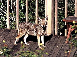
Urban coyotes have little fear of people.
Image by Steve Jurvetson, Menlo Pk, USA;
 CC by 2.0
Do foxes live in North American cities like they do in Europe? It’s true we don’t often read or hear about this, but they do: the city of Mississauga is one municipality that has addressed the issue of foxes in the city. And if those urban foxes have E. multilocularis, that could contribute to the emergence of alveolar echinococcosis in humans. But in North America, we have another canid that likes to live in cities, and it, too, can harbour E. multilocularis: the coyote. A study of urban coyotes in Chicago concluded that there are hundreds, perhaps thousands of coyotes living in that city, and those researchers believe that “the results likely apply to most major metropolitan areas in North America.”

So, while there's no reason to be paranoid about this rare disease, awareness of it isn’t a bad thing. All the usual advice still applies: wash your hands often, keep your dog close, and provide your dog with good veterinary care including screening for intestinal parasites.

Resources


Beaver, Paul C., Jung, Rodney C., and Eddie W. Cupp. Clinical Parasitology 9th ed. Philadelphia: Lea & Febiger, 1984. Pg 534.

City of Mississauga. Animal Services: "Foxes." 1995-2013

ESCCAP. “Worm Control in Dogs and Cats: ESCCAP Guideline 01 Second Edition.” September 2010

Nahorski WL, Knap JP, Pawłowski ZS, Krawczyk M, Polański J, et al. "Human Alveolar Echinococcosis in Poland: 1990–2011." PLoS Negl Trop Dis 2013; 7(1): e1986. doi:10.1371/journal.pntd.0001986

Wagner, Holly. “On the Loose: Urban Coyotes Thrive in North American Cities.” Ohio State Research News. Last Updated 2005.

Wednesday, 26 December 2012

Ivermectin for Bedbugs

Would you take a drug to make yourself poisonous to mosquitoes, or black flies, or wasps? How about taking Ivermectin for bedbugs? I'm not so sure about this - the most obvious problem is that one would have to be bitten before it could work!

Ivermectin for Bedbug Infestation


Bedbug bites can be very uncomfortable.
Taking Ivermectin for bed bugs would
only work if every bug bit at least
once more!
A recent article on Bloomberg.com suggested that giving people oral Ivermectin for bedbugs might be an effective way of dealing with a bedbug infestation. A very small study (three people) found that most bedbugs died if they fed on someone within a day of a dose of Ivermectin, and that 54 hours after the dose, 42% of bugs died after feeding.

Treating people who aren’t sick with drugs has precedent: it’s common for people traveling in places where mosquitoes carry malaria, for example, to take an anti-malarial drug to avoid infection. But while malaria can easily kill you, bedbugs have never been shown to transmit disease to humans.

Ivermectin is an antiparasitic and obviously an insecticide. Like all drugs, it comes with a risk of side effects, some of them quite serious. Would it really make sense to expose large numbers of people – people who aren’t infected with anything - to this drug? It seems to me that treating a dwelling with insecticides is one thing – sometimes not a very good thing – but turning people into insecticide laden bug traps is another.

Bedbug Feeding Habits


Would it even work? Past research has indicated that bedbugs don’t feed every day. A 2009 study indicated that they might feed every two to three days and that they might synchronize their feeding (in other words, the bugs in a colony all tend to feed at the same time). Ivermectin is typically given as a single dose; how would we determine when it’s feeding day for the bedbugs? If the first twenty-four hours is crucial, you’d want to make sure you took the drug on the right day. And what if some survived or didn’t feed that day? And you’d need 100% participation from people staying in the dwelling. Imagine trying to do this in an apartment building. How many doses of Ivermectin would it take?

Finally, I suspect resistance would arise fairly quickly. If 42% of bugs died after feeding at the 54 hour mark, that means 58% survived – and they’d all been exposed to the drug. If their survival was due to them having more natural resistance than the other bugs, and they passed that along to subsequent generations, we’d see more and more resistance.

Before very long, the days of using Ivermectin for bedbugs would be over.


Sources


Gale, J. (2012) “Bed Bugs Dying After Merck Drug Suggests Possible Weapon.” Bloomberg.com

Reinhardt, K., Isaac, D. and Naylor, R. (2010), Estimating the feeding rate of the bedbug Cimex lectularius in an infested room: an inexpensive method and a case study. Medical and Veterinary Entomology, 24: 46–54. doi: 10.1111/j.1365-2915.2009.00847.x

Thursday, 6 September 2012

Thoughts on Naegleria fowleri, "Brain Eating Amoeba"

Naegleria fowleri: a protist that can be a cyst, an amoeba squelching along, or a whirling swimming flagellate; an organism found all over the world that loves warm water, a free living organism that can adopt a parasitic lifestyle; an organism that will almost certainly kill you if it gets into your brain. Beautiful. Fascinating. Deadly.


Naegleria fowleri takes various forms. When it invades a human central
nervous system, it is found as an amoeba or a flagellate.
 Image CDC Image library.

Annual Deaths Due to Naegleria fowleri


Every year during the sweltering days of summer we hear of deaths caused by the “brain eating amoeba.” This year a man died after teaching his daughter how to swim in an Indiana lake, and several children in other American states died after swimming in warm fresh water. Children have died after playing in bath water at home, and the use of neti pots to rinse the sinuses, or ritual inhalation of water into the sinuses, has resulted in deaths as well. The disease is called primary amoebic meningoencephalitis, or PAM.

How Does Naegleria flowleri Infect People?


Naegleria fowleri is just one of more than 20 Naegleria species found in the environment, but to date it is the only one found in human cases of PAM. What’s so special about N. fowleri? Perhaps it has something to do with N. fowleri being a thermophile – in other words it loves warmth. It can survive at temperatures as high as 45ºC, which would make it very comfortable at a normal human body temperature, and impervious to the highest fever. But many of the other species like high temperatures as well, so that’s not the whole answer.

Perhaps it’s important that N. fowleri adapts easily to axenic conditions – meaning that it doesn’t need a community of other organisms around to be happy; it can thrive all by itself. This does make it stand out from the other species, but living inside another organism isn’t exactly axenic, and strains of N. fowleri grown axenically in the lab lose their ability to produce disease. How this characteristic might help it invade the brain in the first place, then, and thrive there, is a tantalizing question – at least to me.

Studies have shown that N. fowleri isolated in the environment contain food vacuoles full of bacteria, whereas those isolated from cases of PAM contain vacuoles full of cell debris. So, when the organism is parasitic, it uses host cells as a food source instead of bacteria. It produces an enzyme that enables it to do this (Chang). This is clearly important, but do we know whether other Naegleria species produce a similar enzyme?

Hot Weather Means Water Sports and Naegleria fowleri


Perhaps it’s a combination of all these factors, and possibly others, that make N. fowleri uniquely equipped to be a “brain eating amoeba.” The question remains to be answered. What’s easier to understand is why it’s so rare, and yet so predictable. In order for N. Fowleri to get into a human brain, very warm water containing the organism must be inhaled into the nasal sinuses. This event is relatively uncommon, but can be expected to happen in the summer months when people – particularly young people – play in the water to cool off.


Sources


Chang SL. “Pathogenisis of Pathogenic Naegleria amoeba.” Folia Parasitol (Praha), 1979; (26)3:195-200.

De Jonckheere JF. “A Century of Reasearch on the Amoeboflagellate Genus Naegleria.” Acta Protozool, 2002; 41: 309-342.

Thursday, 24 May 2012

MOLT: An Internet Game for Diagnosing Malaria

Twenty-four small images of red blood cells appear on the screen. Your job is to click on any that have a malarial parasite inside, removing the image. When you’ve removed all the infected cells, click on “Label all Negative” and another twenty-four cells appear. At the end, you’ll get a score and some information about how many correct choices you made.

You Can Help Diagnose Malaria

Blood films are used to diagnose malaria. The species of Plasmodium
present in the blood can be determined based on the appearance of the
parasites inside red blood cells.
Image: CDC - Public Health Image Library (PHIL) #5942

The game is called MOLT, and it was designed by the Ozcan Research Group at UCLA. Anyone can register and play. The idea is that anyone can be given some basic information about what malarial parasites look like in red blood cells and then be part of an accurate means of correctly diagnosing the disease without having to rely on experts in the field. This would be a huge improvement for malaria diagnosis in parts of the world where malaria kills millions each year and people skilled in diagnosis are rare.

A pilot study of the game using 20 gamers produced results that were within 1.25% of the accuracy of actual experts adept at recognizing malaria, which is pretty impressive. One can imagine an arrangement where someone puts a blood film on a microscope somewhere in Asia or Africa, the images are sent out electronically to potentially millions of gamers around the world, and the answer comes back, positive or negative, in a very short time. If the pilot is any indication, the answer would agree, most of the time, with what an expert would have said.

Crowd-sourcing Games Can Diagnose Malaria and Other Diseases


This has implications for lots of other things that are done by microscopy or other types of imagery: pap smears, fecal smears for parasites, pathology slides etc. It could be improved upon by adding automated scanning techniques and actual experts to the crowd of gamers. These things, plus a larger number of gamers would likely be even more accurate than the gamers used in the pilot. It’s exciting.

I’ve played the game – a number of times. I have lots of experience with reading blood films for malaria, and my biggest issue with the game is that the resolution – the sharpness – of the images is often not good enough

for me to feel completely comfortable with my choices. Platelets sitting on top of red blood cells can look like a parasite. So can debris on the slide. A red cell that’s damaged, or crunched up against another cell, or too darkly stained, or abnormal in some way, etc. etc., doesn’t look like it should to begin with.

I always want to look around a bit, see what the rest of the slide looks like, look for those particular features of a malarial parasite that leave no doubt. In other words, I have a very difficult time deciding whether something is positive or negative on the basis of only one cell (unless the resolution is very good).

My other complaint is with the scoring. I find it ambiguous. When they say “Correct Positive Diagnosis 91%” does that mean 91% of the cells marked as positive were actually positive (false positives), or 91% of positive cases were identified (false negatives). For anyone trying to improve at the game, clarification on this is important.

Play MOLT on BiioGames


Of course I understand that the point is that people who are not experts, and not demanding in terms of excellent microscope optics and parasite features, can still get the right answer if there are enough people providing input. From that perspective, I think the game is brilliant, and I hope it changes the world.

Play the game on Biogames

Read the paper:


Mavandadi S, Dimitrov S, Feng S, Yu F, Sikora U, et al. (2012) Distributed Medical Image Analysis and Diagnosis through Crowd-Sourced Games: A Malaria Case Study. PLoS ONE 7(5): e37245. doi:10.1371/journal.pone.0037245

Friday, 27 April 2012

Do Mosquitoes Transmit Lyme Disease?

While it takes a blood meal, a mosquitoe might
transmit a disease-causing organism. Thankfully,
mosquitoes are not known to transmit Lyme disease.
 Image: US Department of Agriculture
We’ve known for years that Lyme disease is transmitted to humans by ticks. In Europe, it’s usually I. ricinus, the sheep tick, and any of a group of closely related organisms: Borrelia burgdorferi, B. garinii, or B. afzelii; while in my area it’s the deer tick, Ixodes scapularis, and B. burgdorferi. This is enough to worry about as the woods are full of deer and the deer are full of ticks. In some areas more than 30% of deer ticks carry Borrelia, and the ticks are not fussy: they’ll jump onto deer, dogs, cats, and people without hesitation. One hates to think that other biting arthropods could also be transmitting Lyme.

But studies going back as far as the 1980s, and perhaps even farther have found Borrelia in the guts of mosquitoes. Websites devoted to Lyme disease state that mosquitoes are transmitting the disease to humans. Why, then, does the CDC website say “There is no credible evidence that Lyme disease can be transmitted… from the bites of mosquitoes, flies, fleas, or lice” (Lyme Disease Transmission)?

Lyme Disease Organisms (Borrelia) in Ticks


It’s not a matter of a mosquito or tick sucking Borrelia out of one host and then simply injecting it into another like a flying (or crawling) syringe, not like pouring liquid from one glass to another with no change in the contents. Here we are dealing with interactions between living things. Research has shown that things happen in the tick, things that are important in transmission.

In the tick’s gut, Borrelia produces a protein that enables it to persist there for long periods of time, likely aiding survival until the tick feeds again. When the tick is feeding, the spirochete cuts back on this protein and produces a different one instead, one that enables it to invade the tick’s salivary gland and then be transmitted to the new host in the tick’s saliva.

Similarly, Borrelia is able to enhance a tick protein that protects both tick and spirochete from attack by the host immune system: “Borrelia burgdorferi, the Lyme disease agent, is critically dependent on the presence of the tick protein Salp15 when infecting the host” (Schwalie and Schultz). The extended time that a tick spends feeding (days) provides plenty of time for this interaction to take place.

Lyme Disease Organisms (Borrelia) in Mosquitoes


In contrast, while Borrelia has been detected in mosquito guts and saliva, it doesn’t appear to survive there very long, probably because the proteins that support it in ticks don’t work in mosquitoes. Salp15, too, is a tick protein that won’t be available to help out in a mosquito, and mosquitoes take only minutes to obtain a blood meal, compared to days for a tick. Put simply, mosquitoes are not competent vectors of B. burgdorferi; they just don’t have the right stuff. While it’s not impossible that a mosquito bite could contain the spirochetes, it’s unlikely, and it’s even more unlikely Borrelia would succeed in setting up an infection. Mosquitoes are not significant vectors of Lyme disease.


References

Fontaine et al: Implication of haematophagous arthropod salivary proteins in host-vector interactions. Parasites & Vectors 2011 4:187 doi:10.1186/1756-3305-4-187

Hovius, JWR. Tick-host-pathogen interactions in Lyme borreliosis. Dissertation, Academic Medical Center, University of Amsterdam 2009

Kosik-Bogacka D, Bukowska K, Ku?na-Grygiel W. Detection of Borrelia burgdorferi sensu lato in mosquitoes (Culicidae) in recreational areas of the city of Szczecin. Annals of Agricultural and Environmental Medicine 2002, 9, 55–57

Schwalie PC, Schultz J.  Positive Selection in Tick Saliva Proteins of the Salp15 Family. Journal of Molecular Evolution Volume 68, Number 2 (2009), 186-191, DOI: 10.1007/s00239-008-9194-1

Magnarelli LA, Anderson JF. Ticks and Biting Insects Infected with the Etiologic Agent of Lyme Disease, Borrelia burgdorferi. Journal of Clinical Microbiology Aug. 1988, p. 1482-1486

Wednesday, 7 March 2012

Toola, Sea Otters, and Toxoplasma gondii

Reports in March 2012 of the death of Toola, a Toxoplasma gondii-infected sea otter who lived out her days at the Monterey Bay Aquarium, reminded me of the threat that T. gondii poses to marine mammals. Toola suffered from neurological damage thought to have been caused by the parasite and required daily anti-seizure medication. Among other things, she was the poster otter for legislation and other efforts to protect marine mammals from various health risks. And she was cute too.

Sea otters frequent the California coast, where they may
become infected with T. gondii. The consequences can be deadly.
Image by Mike Baird, Morro Bay, USA. CC BY 2.0

How do Sea Otters Get Toxoplasma gondii?


My impression has been that the risk of acquiring T. gondii has been rising in marine mammals, and that this is likely to be the result of runoff – oocysts being washed off the land into coastal waters. This made sense to me when considering the number of feral and roaming domestic cats, and the quantity of cat feces that must be carried into coastal waters by runoff (this has actually been studied: “domestic feline faecal deposition in communities adjacent to Estero Bay was conservatively estimated at 107 metric tonnes/year, or 26 kg/ha:” Miller et al.) I was surprised; therefore, to read that the majority of California sea otters tested in the 2008 study reported by Miller et al had a unique strain (dubbed Type X) that is not typically found in domestic cats.

Rather, the paper by Miller et al. reports that Type X T. gondii was found in wild felids (mountain lion, bobcat) and foxes. While foxes might be doing relatively well in urban areas, the number of wild felids is down from what it must have been before humans covered the west coast of North America with concrete and asphalt. So if domestic cats aren’t to blame, why are there more infected marine mammals now than before?

Humans and Mollusks Spread Toxoplasma to Marine Mammals


One answer apparently lies in all that concrete and asphalt. Hardscaping of the coast reduces the amount of runoff that’s absorbed into the ground before it spills into the sea. In addition:

  • Human development has reduced wetlands, which provide natural filtration for runoff.

  • Bivalves such as mussels flourish near storm sewers and have been shown to filter organisms, including T. gondii oocycts out of the water and concentrate them in tissue.

  • Sea otters feed on mussels and other bivalves, consuming at least 76 mussels each day.

Studies done on land mammals have shown that a single oocyst can potentially be the source of chronic toxoplasmosis. Given those odds, its not surprising that Toola, and lots of other California sea otters (and other marine mammals) are infected with T. gondii.

Read the paper:

Miller, M.A., W. A. Miller, P. A. Conrad et al. "Type X Toxoplasma gondii in a wild mussel and terrestrial carnivores from coastal California: New linkages between terrestrial mammals, runoff and toxoplasmosis of sea otters." International Journal for Parasitology: 38(11), 2008

Monday, 30 January 2012

Sanitation Prevents Intestinal Worm Infections

A pit latrine in Haiti. Even a simple design
such as this will do much to reduce
contamination of the environment, and result
in fewer intestinal parasites.
Image by Rémi Kaupp. CC BY-SA 3.0
A paper in PLOS Medicine (January 24, 2012) reports that “sanitation is associated with a reduced risk of transmission of helminthiases to humans.” The authors looked at 36 previously published studies that measured prevalence of intestinal helminths (A. lumbricoides, large intestinal roundworm; T. trichiura, whipworm; and hookworm) compared to availability and use of sanitary facilities. They found that “people who either had or used a latrine were half as likely to be infected with a soil-transmitted helminth as people who neither had nor used a latrine.”

Contaminated Soil and Intestinal Worms


I submit that there are no surprises here. One acquires hookworm by coming in contact with hookworm larvae from feces contaminating the soil. They penetrate skin. Trichuris trichiura and A. lumbricoides eggs, infective a week or so after being deposited in warm moist soil in feces, must be swallowed. Obviously if feces were deposited in a pit latrine, septic system or other sanitary arrangement, instead of on the ground, those eggs and larvae would not be available to infect new hosts.

The fact that intestinal helminthes are much less common, even rare, in developed countries is no mere accident of climate, especially for the tough A. lumbricoides. It is because the majority of people in developed countries don’t defecate outside on the ground.

Parasite Prevention: Sanitation Works


The best point in this paper, though understated, is that periodically treating people for intestinal worms is perhaps not the best long term approach to getting rid of these parasites. Without good sanitation, people will quickly be reinfected due to contamination of their environment. Lets build toilets.

Read the paper:

Ziegelbauer K, Speich B, Mäusezahl D, Bos R, Keiser J, et al. (2012) "Effect of Sanitation on Soil-Transmitted Helminth Infection: Systematic Review and Meta-Analysis." PLoS Med 9(1): e1001162. doi:10.1371/journal.pmed.1001162

Monday, 9 January 2012

Helping Mosquitoes Fight Off Malaria

Anybody who knows anything about malaria knows that one catches it from a mosquito bite. Mosquitoes don’t just physically carry the parasite from person to person: they are a required host for Plasmodium spp., the agents of malaria. In the mosquito, the parasites multiply sexually, producing tiny forms called sporozoites which are injected into the next person the mosquito bites.

[caption id="attachment_382" align="alignleft" width="300" caption="Anopheles stephensi feeding; CDC, public domain image"]Anopheles stephensi transmits malaria[/caption]

Clearly, the mosquitoes are infected just as people are, but we seldom feel sorry for the poor mosquitoes because, well, we hate them for all sorts of reasons. The mosquito, however, does have an immune system which tries to fight off invading Plasmodium sp. parasites; Mosquitoes don’t mean to transmit these dangerous parasites.

Humans have had a long and costly battle with malaria which, so far, we have not won. Though not self-evident perhaps, it makes sense that we might be able to enlist the help of the lowly mosquito to our mutual benefit, and that’s what some researchers at Johns Hopkins University have done. Yuemei Dong et al. have genetically modified the immune system of a mosquito species, Anopheles stephensi, giving it an enhanced ability to fight off invading Plasmodium falciparum, the worst of the malaria parasites in humans.

In order for this research to prove useful in the real world, the modified mosquitoes would have to be released into the wild and allowed to breed with wild populations (and hopefully do better than the wild type). Aside from the obvious need for caution when releasing a genetically modified organism into the wild, at this point we still don’t know whether:

  • the resistant mosquitoes will do as well in the wild, faced with different A. falciparum strains

  • other Anopheles spp., also malaria vectors, can be similarly modified (there are about 40)

  • Plasmodium falciparum will develop resistance to the mosquito resistance

  • all other species of Plasmodium infecting humans can be targeted this way


This breakthrough is not the answer to the battle against malaria yet, but it may be part of the answer.

Read the paper:

Dong Y , Das S , Cirimotich C , Souza-Neto JA , McLean KJ , et al. 2011 “Engineered Anopheles Immunity to Plasmodium Infection” PLoS Pathog 7(12): e1002458. doi:10.1371/journal.ppat.1002458